
Arthrogram Imaging Services

What is an arthrogram? | What joints can be examined? | MRI and CT arthrograms |Preparation | What to expect | After your arthrogram | Understanding your results


An arthrogram is a specialized imaging exam used to evaluate joint pain, injuries, and abnormalities. By injecting a small amount of contrast material directly into a joint, an arthrogram allows your radiologist to see structures that may be difficult to evaluate with standard imaging alone.

Arthrograms are commonly performed in combination with MRI or CT imaging to provide detailed pictures of cartilage, ligaments, tendons, and other structures within a joint. These images help your healthcare provider identify potential causes of discomfort and determine appropriate treatment options.
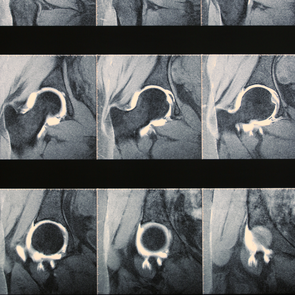
arthrogram

What is an arthrogram?

Arthrography is a diagnostic imaging procedure that provides a detailed look inside a joint. During the exam, contrast material is injected into the joint space using imaging guidance. The contrast helps outline important structures, making subtle injuries or abnormalities easier to identify.

An arthrogram may be recommended when traditional imaging does not provide enough information to explain persistent joint pain, limited mobility, instability, or other symptoms.

Arthrograms can help evaluate conditions such as:

  • Cartilage and labral tears – Damage to the cartilage or labrum that helps support and stabilize a joint.
  • Ligament and tendon injuries – Tears or abnormalities affecting the tissues that connect and support bones and muscles.
  • Joint instability – Structural problems that may contribute to repeated dislocations or a feeling that a joint is unstable.
  • Persistent joint pain – Unexplained discomfort that may require a closer examination of the joint’s internal structures.

What joints can be examined?

Arthrograms are commonly used to evaluate the shoulder and hip, but they may also be performed on the knee, wrist, elbow, or ankle.

Your healthcare provider will determine whether an arthrogram is appropriate based on your symptoms, medical history, and previous imaging results.

MRI and CT arthrograms

Depending on the area being examined and the suspected condition, your healthcare provider may recommend an MRI or CT arthrogram.

MRI Arthrogram – Combines an injection of contrast material with magnetic resonance imaging (MRI). This technique provides detailed images of soft tissues, including cartilage, ligaments, tendons, and the labrum. MRI does not use ionizing radiation.

CT Arthrogram – Combines contrast material with computed tomography (CT) imaging to produce detailed cross-sectional images of a joint. CT arthrograms may be recommended when detailed evaluation of certain joint structures is needed or when MRI is not suitable.
Both examinations can provide valuable information to help your healthcare provider diagnose joint conditions and plan treatment.

Preparing for your arthrogram

Arthrograms generally require minimal preparation. Our team will provide any specific instructions before your appointment based on the type of examination being performed.

To help prepare for your visit:

  • Wear comfortable, loose-fitting clothing and leave jewelry and other valuables at home.
  • Inform our team of any allergies, particularly previous reactions to imaging contrast material.
  • Provide a list of medications you take, including prescription medications, over-the-counter products, and supplements.
  • Tell our team about any medical conditions, including kidney problems, bleeding disorders, or recent illnesses.
  • Inform your technologist if you are pregnant or think you might be pregnant.
  • If your examination includes MRI, let our team know about any implanted medical devices, metal implants, or other metal in your body.

Most patients can eat and drink normally before an arthrogram unless instructed otherwise. Do not stop taking prescribed medications unless directed by your healthcare provider.

What to expect during your arthrogram

An arthrogram typically involves two steps: injecting contrast material into the joint and obtaining detailed images of the area.
You will be positioned on an examination table, and the skin around the joint will be cleaned. A local anesthetic is generally used to numb the area and minimize discomfort.

Using imaging guidance, such as fluoroscopy (real-time X-ray imaging), a radiologist or other qualified provider carefully places a thin needle into the joint space and injects the contrast material. Imaging guidance helps ensure accurate placement.

You may experience a brief sensation of pressure, fullness, or mild discomfort as the contrast enters the joint. The needle is then removed, and you may be asked to gently move the joint to help distribute the contrast.

If your arthrogram is being performed with MRI or CT, additional images will be obtained following the injection.

The total appointment time varies depending on the joint being examined and the type of imaging required. Our team will guide you through each step and help keep you comfortable throughout your visit.

After your arthrogram

Most patients can return home shortly after their examination and resume many of their normal daily activities.

It is common to experience mild soreness, swelling, or a feeling of fullness in the joint following the contrast injection. These symptoms are generally temporary and typically improve within 24 to 48 hours.

Your healthcare team may recommend:

  • Avoiding strenuous exercise or heavy use of the affected joint for at least 24 hours.
  • Applying a cold compress to help relieve temporary soreness or swelling.
  • Following any specific activity restrictions or care instructions provided after your examination.

Although complications are uncommon, contact your healthcare provider if you experience increasing pain, significant swelling, redness, fever, or other concerning symptoms following your procedure.

Understanding your results

After your arthrogram, a radiologist will carefully review the images to evaluate the joint and identify any abnormalities.

A detailed report will be sent to your referring healthcare provider, who will discuss the findings with you and recommend any appropriate next steps.

Arthrogram imaging can provide valuable information when evaluating joint injuries and unexplained discomfort, helping your healthcare team develop a treatment plan tailored to your needs.

Please contact our imaging team at (360) 255-6330 with any questions or concerns.