Medical Imaging Services
Looking for answers to your medical diagnostic imaging questions? We can help!
Bellingham Advanced Medical Imaging is here to give you and your doctor the answers you need to make the best decisions about your health care. And, complete clarity about the various types of medical imaging available.
Our team of board-certified radiologists, technologists and support staff knows that this can be a stressful time for you. You want and deserve answers as soon as possible.
We use advanced medical imaging technology to help you and your doctor can get the answers you need quickly and safely. We do this by making sure you’re at the center of everything we do at Bellingham Advanced Medical Imaging.
Learn more about the different kinds of medical imaging exams we have at Bellingham Advanced Medical Imaging
CT Scan | MRI Imaging | Ultrasound | X-Ray | Fluoroscopy | DEXA Scan
Ultrasound
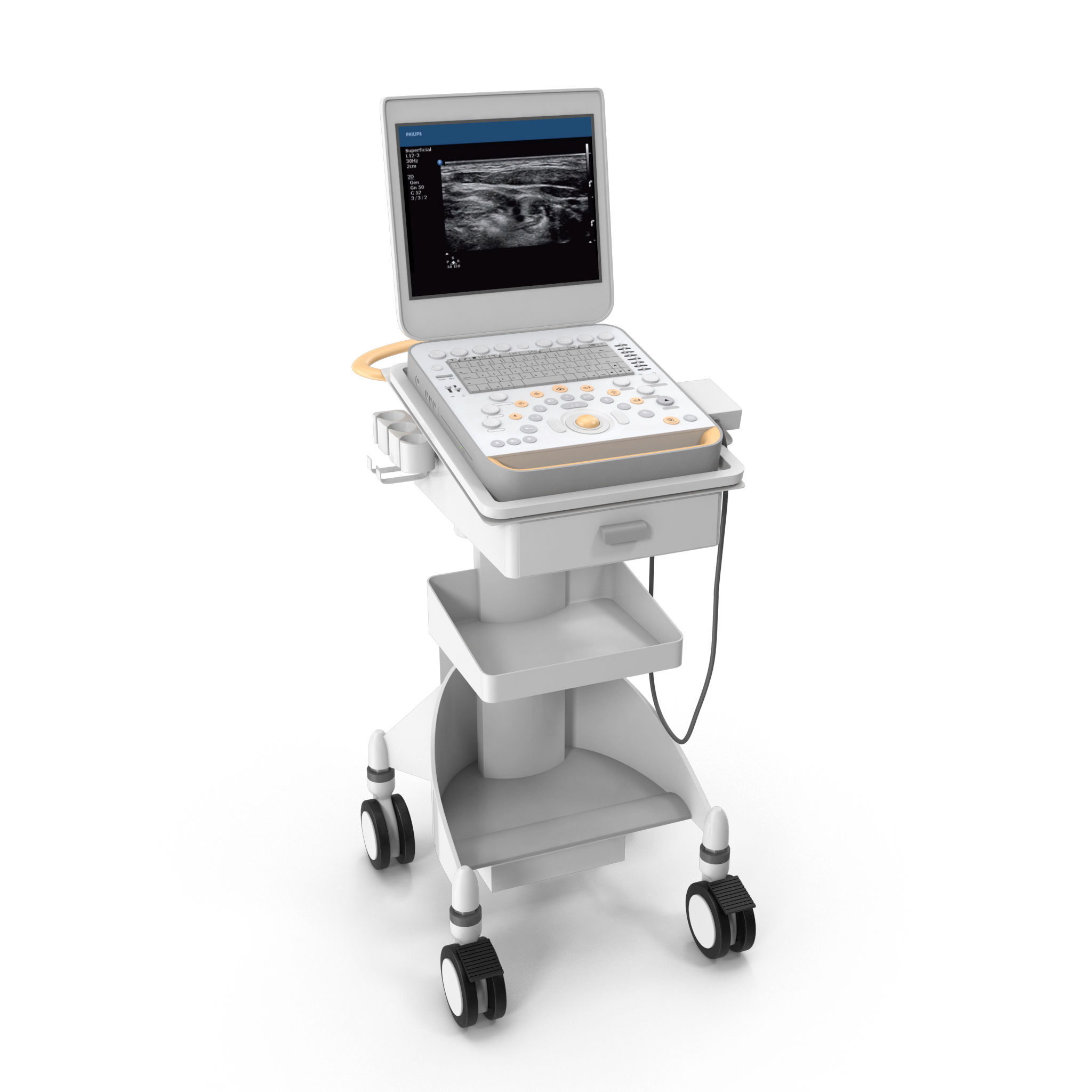
Ultrasound uses sound waves to create images of your organs, blood vessels, and other structures inside your body. Because ultrasound is fast and does not use ionizing radiation, it is relatively safe. This makes ultrasound a preferred imaging option for children and pregnant patients.
High-Frequency Sound Waves - Ultrasound uses a small tool called a transducer to send high-frequency sound waves into an area of your body. The transducer also receives the sound waves as they bounce back. The ultrasound machine interprets those sound wave echoes to create an image of your internal organs and tissues. Ultrasound images are obtained in real-time, which allows the evaluation of dynamic processes such as heart contraction, blood flow, and stomach emptying.
Doppler Ultrasound - This type of ultrasound measures the change in pitch of the ultrasound waves with motion, which enables the evaluation of blood flowing in veins and arteries. Doppler ultrasound can also be used to check for heart motion very early in pregnancy.
For additional information about specific tests, please follow the links below:
Abdominal Ultrasound
Carotid Ultrasound
Obstetrical Ultrasound
Pelvic Ultrasound
Scrotum Ultrasound
Thyroid Ultrasound
Vascular Ultrasound
Venous Ultrasound
X-Ray
Painless and fairly quick, X-rays give you and your health care provider a look inside your body. They are a simple, non-invasive, and quick way to help spot broken bones, foreign objects, and even diseases in certain parts of your body.
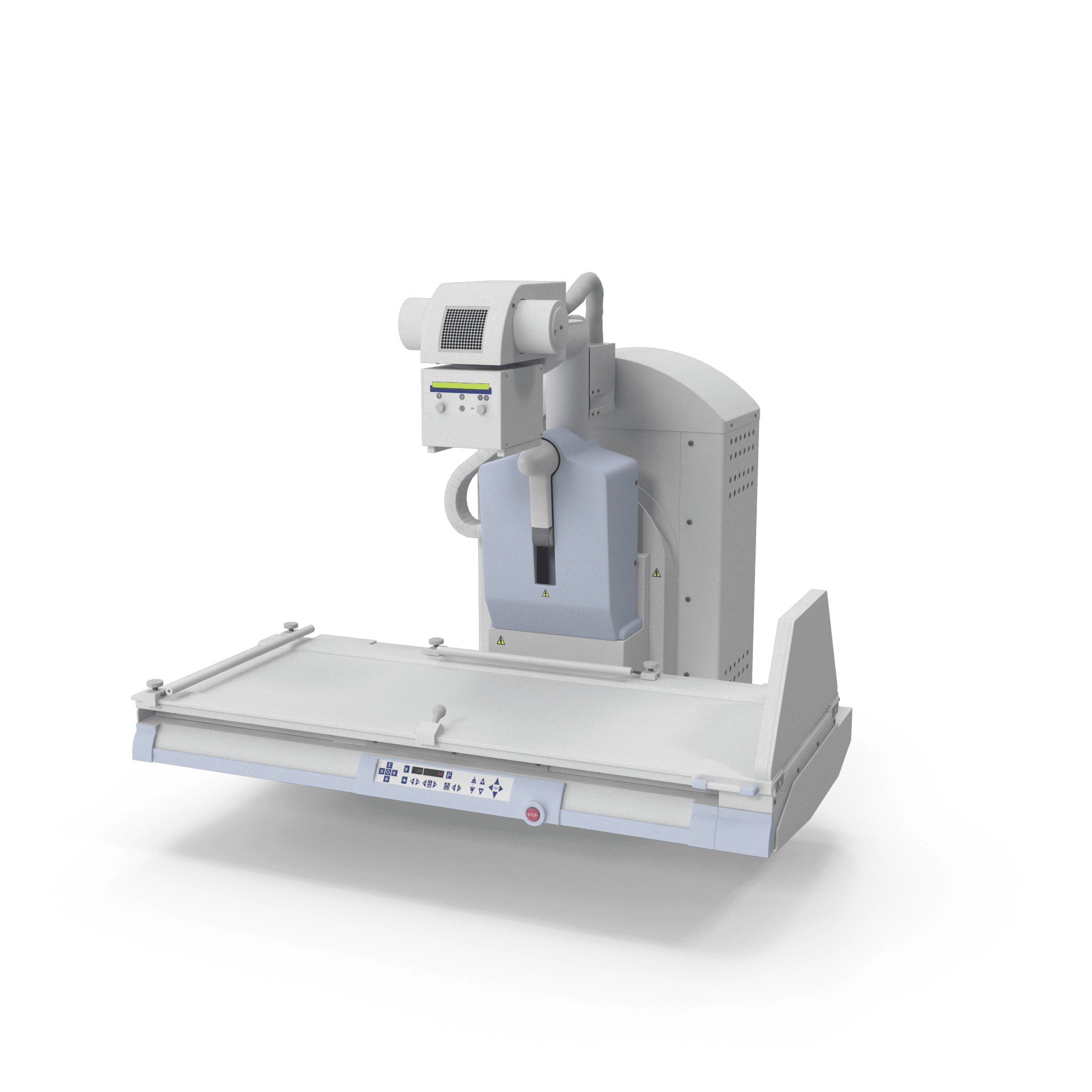
Dense and Soft Tissue - X-rays use a small amount of radiation to provide images of your body. As the radiation passes through, denser structures (like bones) block the radiation, while soft tissue (like the lungs) does not. A digital X-ray sensor captures the X-rays that pass through, leaving an image that can show abnormalities such as bone fractures, foreign objects, and pneumonia.
Fluoroscopy
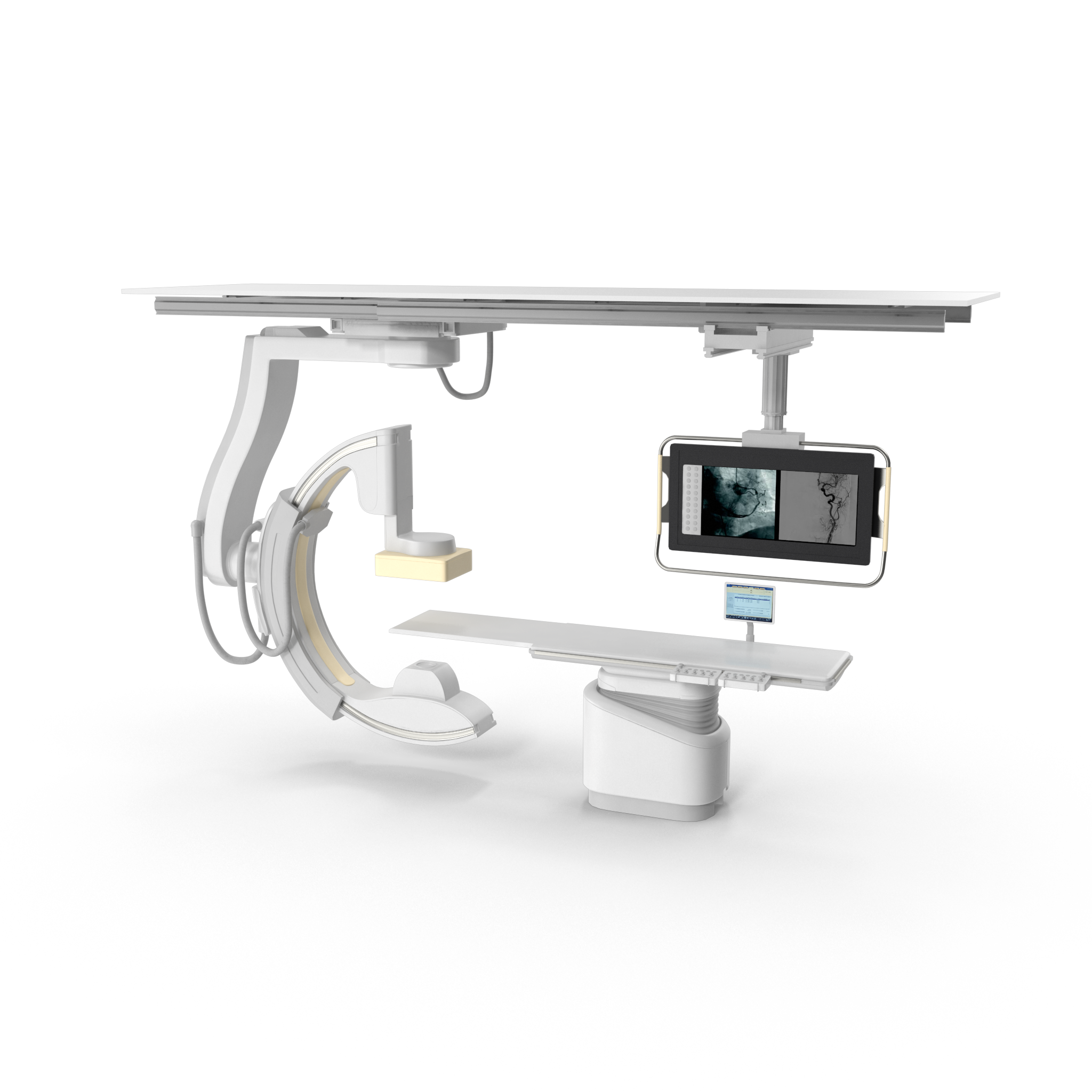
While standard X-ray takes still images, fluoroscopy allows real-time imaging with live X-rays. At Bellingham Advanced Medical Imaging, we primarily use fluoroscopy when injecting contrast for patients receiving an MRI or CT scan, this procedure is known as arthrography.
DEXA Scan
Bone Densitometry - also called dual-energy x-ray absorptiometry, DEXA, or DXA, is a type of radiology that uses a very small dose of ionizing radiation to produce pictures of the inside of the body, lower spine, and hips. Specifically, to measure bone loss. A DEXA scan is most commonly used in the diagnosis of osteoporosis as well as to assess an individual's risk for developing osteoporotic fractures. A DEXA scan is simple, quick, and noninvasive. It's the most commonly used standard method for diagnosing osteoporosis.
A DEXA exam requires little to no special preparation. Tell your doctor and the technologist if there is a possibility you are pregnant or if you recently had a barium exam or received an injection of contrast material for a CT or radioisotope scan. Leave jewelry at home and wear loose, comfortable clothing. You may be asked to wear a gown. You should not take calcium supplements for at least 24 hours before your exam.
NOTE: The information contained within this website should not be considered medical advice and is not intended to replace the consultation of one of our qualified radiologists. Skagit Radiology has compiled this information to the best of its ability; however, it is possible there may be more current information following the posting of this information. Please consult your physician and/or radiologist for information about your particular situation.
